
 Abdominal Ultrasound:
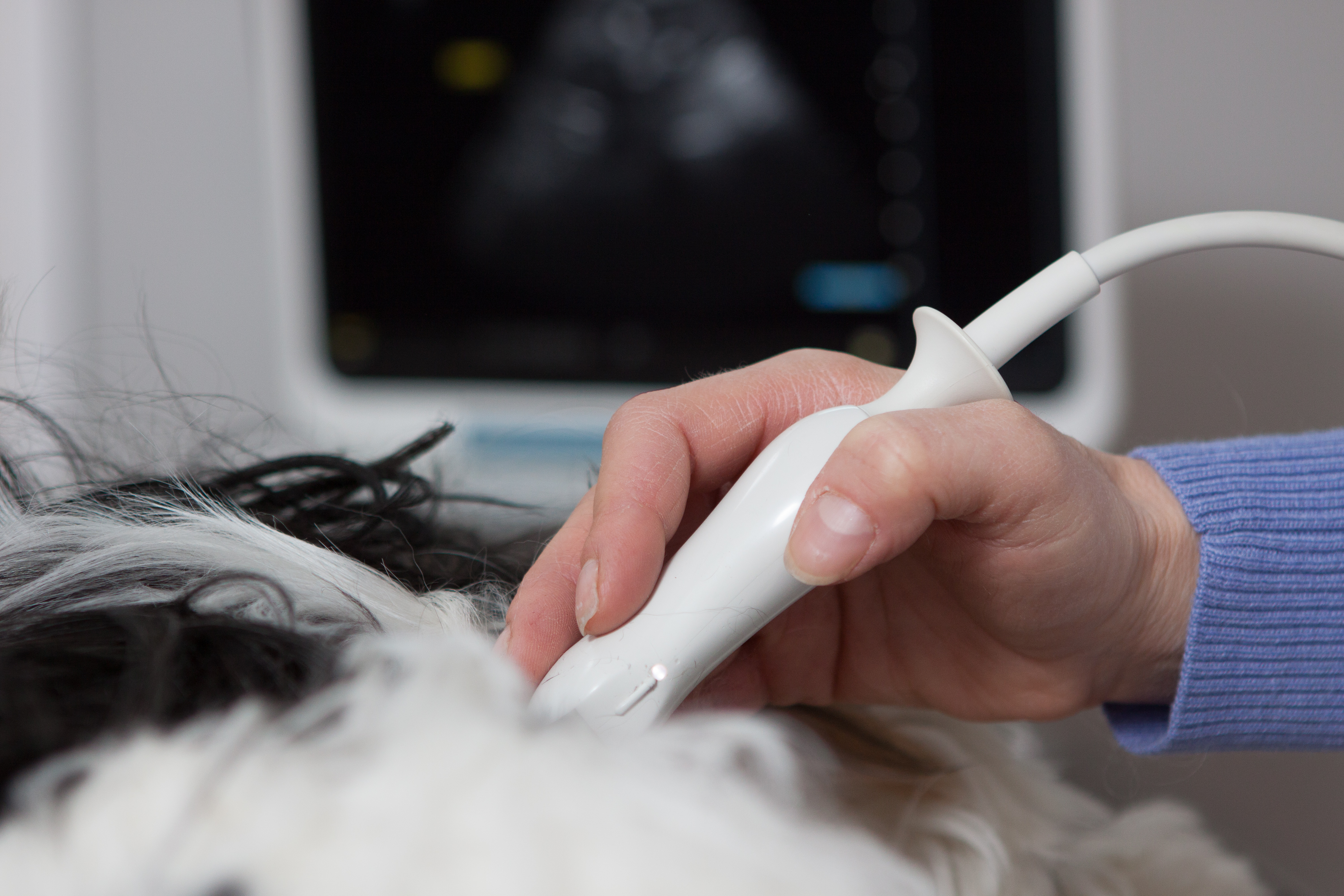
Abdominal ultrasound is a staple as a non-invasive diagnostic test in the workup of our patients. A complete ultrasound of the abdomen is performed during each exam as not to miss occult or concurrent disease. On all exams, interpretation is made cage side with a verbal and written report generated at the time of the exam. A fully-typed written report will be provided within 24-48 hours and will be sent by email or fax by one of our experienced transcriptionists, Kristin or Sarah. Images will be saved digitally to thumb drives (provided by me and left at your clinic for future cases). Digital images can be easily transmitted to referral clinics as needed and can be integrated into digital medical records depending on the hospital’s computer system.
An ultrasound of the abdomen involves evaluating all the abdominal contents including the liver, gall bladder, spleen, kidneys, adrenal glands, urinary bladder, stomach, intestines, colon, pancreas, and lymph nodes. Evaluating for the location and origination of masses and causes of ascites are routine as well. Pulmonary infiltrates can often be seen across the diaphragm, which may prompt further investigation if pulmonary disease is suspected. Dr. Frank is also skilled in advanced sonographic procedures such as detecting the presence of portosystemic shunts.
Echocardiography:
Echocardiography is performed to evaluate the structure and function of the heart in both acquired and congenital disease processes. Each exam includes:
- Two-dimensional views with assessment of chamber sizes and valve structure
- M-mode exam with normalization of cardiac parameters
- Color flow and spectral flow Doppler of all four valves
- Diagnosis of pericardial fluid, right atrial masses and heart based masses; pericardiocentesis and therapeutic drainage
- Sonographic assessment of the pulmonary parenchyma if indicated
- Review of thoracic radiographs, ECG and blood pressure as available
- Doppler blood pressure measurement if needed or requested
Clinical recommendations are made at the time of the exam. Follow-up echocardiograms may be recommended depending on the case. Telephone consultation for aid in long-term case management will be provided upon request.
Some cases will ultimately require referral to a local board-certified cardiologist and this will be recommended as necessary. In the event of congenital heart disease, Dr. Frank will be able to help guide you if an interventional procedure is available or necessary for your patient and would require referral.
Thoracic Ultrasound:
Ultrasound of the chest is not limited to the cardiac exam. In general, evaluation of the lungs is best accomplished via thoracic radiographs or CT scanning. However, pulmonary infiltrates can often be detected with ultrasound and may lead to additional diagnostics. Pulmonary edema can be detected with ultrasound although thoracic radiographs may be subsequently recommended. Small diffuse metastatic lesions can often be visualized.
- Assessment and sampling of pulmonary/intra-thoracic masses is possible when the mass is in close contact with the thoracic wall Masses can also be detected when they are anatomically located adjacent to the heart and diaphragm
- Diagnosis and confirmation of diaphragmatic hernia and pericardial- peritoneal diaphragmatic hernia
- Thoracocentesis for diagnostic sampling and therapeutic drainage
Neck: Thyroid and parathyroid glands
Advanced ultrasound techniques include the ability to visualize the thyroid and parathyroid glands, which can be important in the work up of hypercalcemic patients, or in patients with a suspected thyroid mass.
Internal Medicine Consultations
Dr. Frank is a board certified internal medicine specialist with the American College of Veterinary Medicine (ACVIM). In each ultrasound consultation, she draws on her medical expertise during the exam as well as in her recommendations for future diagnostics and therapy. However, upon request. Dr. Frank can provide a more extensive internal medicine consultation with complete review of the case history and chart, full examination of the patient, and a more extensive case report. Consultations can be done in conjunction with an ultrasound or separately, although ultrasound is often used in an internal medicine work up. Internal medicine consults are billed separately and must be booked in advance.
Additional Diagnostics: Bone Marrow Biopsy and Joint Taps
Bone marrow biopsy (aspirate and core biopsy) is an important diagnostic tool in the work up of cases of blood disorders, such as cytopenias (anemia, leukopenia, pancytopenia) or proliferative bone marrow disorders (e.g. severe lymphocytosis). This is also commonly performed in the work up or staging of various cancers such as lymphoma/leukemia and multiple myeloma.
Joint taps can be performed and are indicated in the work up of polyarthritis.